
 |
 |
 |
 |
 |
 |
 |
 |
 |
 |
 |
 |
 |
 |
 |
 |
 |
 |
 |
 |
 |
 |
 |
 |
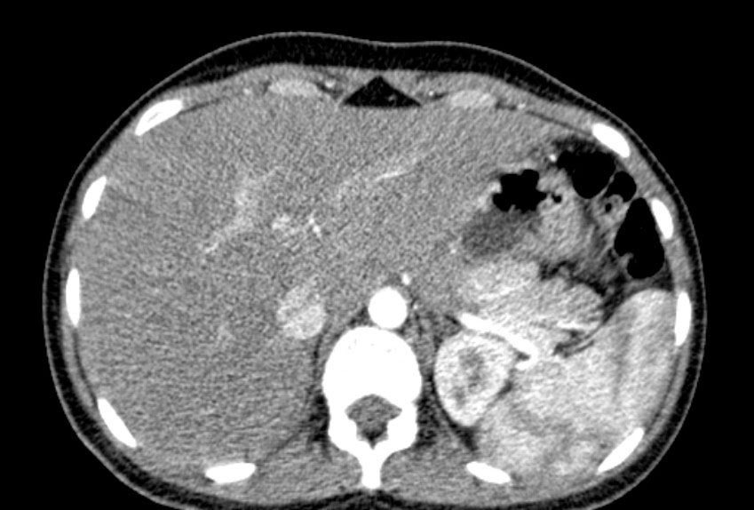
Here the papillary process is seen as an exophytic projection, seen extending into the perigastric region.
In the axial sections it appears as a separate 'lesion', in close relation to tail of pancreas.
In all imaging series, it is seen isoattenuating to the liver parenchyma.
CT for this young female patient was done for suspicion of appendicitis and renal calculi.
"The papillary process of the caudate lobe of the liver may appear separate from the liver and thus mimic lymph nodes or a pancreatic mass." (Radiology. 1989 Dec;173(3):631-3. Papillary process of the caudate lobe of the liver: sonographic appearance. Donoso L1, Martínez-Noguera A, Zidan A, Lora F.)
No comments:
Post a Comment