Friday, August 15, 2014

Active arterial extravasation in Blunt Abdominal Trauma


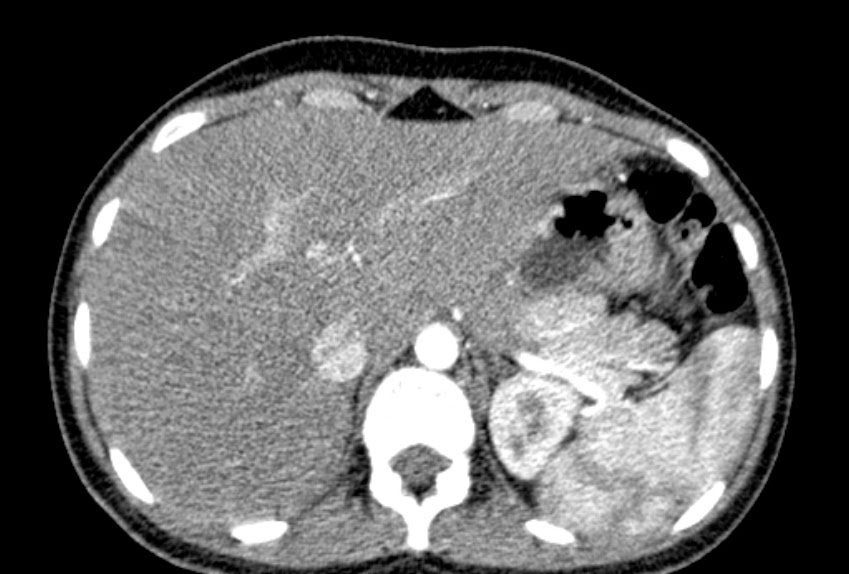
H/o Road Traffic Accident, Middle aged male, with maintained SpO2 and Blood Pressure.

The following plain CT sections show Hematoma involving right diaphragmatic crus with marked enlargement compared to the contralateral side. Hyperdensities s/o blood clots are noted within the hematoma. Mild amount of bilateral retroperitoneal hemorrhage was seen, with associated mild perinephric hemorrhage.


The following 3 images, shows the serial axial section during PLAIN / Non-contrast CT, Arterial Phase (25sec) and during Portal Venous Phase (60secs) respectively. Images demonstrate small area of contrast extravasation from the lateral margin of abdominal aorta, with minimal increase in the portal phase --> s/o an Active Arterial Hemorrhage.


These serial 5 images that follow, shows pooling of contrast in the 5 minute delayed images. Areas of contrast pooling are marked with large red arrows.


There was also a small anterior cortical laceration of right kidney.


Pseudolesion : Papillary Process of Caudate Lobe


Here the papillary process is seen as an exophytic projection, seen extending into the perigastric region.
In the axial sections it appears as a separate 'lesion', in close relation to tail of pancreas.

In all imaging series, it is seen isoattenuating to the liver parenchyma.

CT for this young female patient was done for suspicion of appendicitis and renal calculi.

"The papillary process of the caudate lobe of the liver may appear separate from the liver and thus mimic lymph nodes or a pancreatic mass."  (Radiology. 1989 Dec;173(3):631-3. Papillary process of the caudate lobe of the liver: sonographic appearance. Donoso L1, Martínez-Noguera A, Zidan A, Lora F.)


Case of the Day : Sclerotic Sphenoid lesion

Sclerotic Sphenoid Lesion
Patient is a middle aged female with headache.


DIAGNOSIS : FIBROUS DYSPLASIA of Sphenoid bone (right).

Sclerotic lesion with the characteristic / diagnostic 'Ground Glass matrix' is seen here involving the sphenoid body and  lesser wing mainly of the right side. Lesion is completely involving the right optic canal, with no narrowing of the canal.

Polyostotic Fibrous Dysplasia - is a feature of McCune Albright Syndrome - associated with precocious puberty and pigmentation. MA-Syndrome usually have Cafe-au-lait patches, which is described to have a 'Coast of Maine' appearance - irregular margins (c.f 'Coast of California' appearance of Cafe-au-lait patches of neurofibromatosis).

Polyostotic fibrous dysplasia having a rare association of intramuscular myxomas is seen in Mazabraud Syndrome.

Saturday, August 9, 2014

Castellvi Type IIIb LSTV


Castellvi type IIIb Lumbo-Sacral Transtional vertebra

Bilateral osseous  fusion of the enlarged L5 transverse processes with sacral ala (Sacralization of L5 vertebra) is seen here.

See description of various types of LSTV : Castellvi types


LinkWithin

Related Posts Plugin for WordPress, Blogger...