Wednesday, November 29, 2017

Aortic Dissection : Stanford Type B


Patients with abdominal pain often undergoes CT evaluation for further assessment after a baseline USG. Diagnosing aortic dissection is easy in post contrast CT (Angiogram) images, but when the patient comes for CT Abdomen, reasonable observation and high degree of suspicion is needed, to proceed with CT Chest and Abdomen Angiogram (rather than abdomen alone), otherwise we would end up taking multiphase images of abdomen alone. The therapeutic decision in dissection depend upon the detecting the involvement of preductal aorta (Stanford A), which requires emergency surgical management. Thus if we fail to detect / suspect dissection in plain study, we have to take repeat angiogram of the thoracic aorta later.

The following is a case of a 40yr old female patient who presented with abdominal pain radiating to back. USG did show slightly prominent aorta for age, which is actually a good pickup, since there was no other abnormality in abdomen.

CT Abdomen was requested for further evaluation of the abdominal pain. In the plain CT sections, the aorta was seen dilated, with proximal abdominal aorta measuring up to ~3.5cm in outer diameter. Highest section of the scan did show the dilated distal descending thoracic aorta with hyperdense peripheral crescent, indicating acute intramural hematoma (/thrombosed false lumen of dissection), which necessitates the scan to be taken from arch vessels to iliac arteries, to assess the involvement of ascending aorta, arch and to look for the inferior extent.


Dilated proximal abdominal aorta, raising the suspicion of dissection in a 40yr old female patient.


Hyperdense crescent indicating acute intramural hematoma.


Above image shows the proximal end of the intramural hematoma, beginning just distal to the left subclavian artery origin, making it a Stanford type B dissection. Ascending aorta and arch branches were not affected (not shown).


The hyperdense hematoma can be seen slightly rotating / whirling in an anticlockwise fashion, to come towards the left lateral aspect of aorta (see below), from the posteromedial aspect.


The dissection flap was only visualized from the D11 vertebral level.


The above image showing relationship of the true lumen (green arrow) and false lumen (orange arrow). As going distally the false lumen is seen enlarging in size.


3D-VRT images showing the dissection flap (yellow arrows).


It's important to determine the origins of branch vessels before endograft or stent placement to avoid end organ ischemia.


Axial sections showing the origins of major abdominal aortic branches, with true lumen on right and false lumen on left side. Celiac trunk, left renal artery and IMA are seen arising from the false lumen.


Dissection is the spontaneous longitudinal separation of intimal and adventitial layers because of blood traversing and splitting through the media. The inciting factor is due to an intimal tear, which allows the blood to enter into the media. The additional lumen thus formed within the media is called as the false lumen.

Aortic dissection is divided into two types according to the Stanford classification. If ascending aorta is involved then its termed type A and if it involves aorta distal to the left subclavian artery alone then its termed type B dissection.

The high attenuation within the aortic wall is either due to acute intramural hematoma or due to hyperdense false lumen.

Sometimes within the hyperdense false lumen, residual flow can be seen (as shown below).


Residual flow channel in false lumen (Orange arrow).


References:
1. AJR 2001 July article.
2. RG March-April 2010 article.

Leaking Abdominal Aortic Aneurysm (AAA) in plain CT


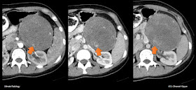
Elderly male patient with abdominal pain, with raised Serum Creatinine underwent Plain CT Abdomen, which showed a large fusiform aneurysm of the abdominal aorta, measuring ~12cm in length and 6cm in maximal diameter. Left retroperitoneal massive hemorrhage is noted lifting the left kidney anteriorly (above image).


Aneurysm (orange arrows) and the surrounding hemorrhage (yellow arrows).


Ogilvie Syndrome or Acute Colonic Pseudo Obstruction

Ogilvie Syndrome

Also known as Acute Colonic Pseudo Obstruction, Ogilvie syndrome is a potentially fatal clinico-radiological conditon with features akin to large bowel obstruction without any mechanical obstruction. 

It is most commonly seen in elderly and usually unwell patients, with multiple probable or causative factors like electrolyte imbalances, diabetes mellitus, uremia (renal failure), recent surgery, medications (eg: Opioids) etc. Pathological analysis of resected specimens showed reduced intramural ganglion cells in most cases.

Even though there is no mechanical obstruction  this condition can lead to bowel gangrene and subsequent peritonitis, increasing the mortality risk. Therefore early identification of this entity and nasogastric suction, medical management (anticholinesterases), decompression by rectal tube, colonoscopic exsufflation is needed. Sometimes cecostomy under fluroscopic guidance and rarely cecectomy might be required.

Patients usually present with abdominal pain and distension with preserved bowel sounds on auscultation.

A plain abdominal radiograph usually shows dilated colonic loops (presence of haustra), more often the right colon and transverse colon, with relative lack of air fluid levels (which are more commonly seen with SBO). Rectal gas is usually seen in radiographs, indicating lack of mechanical obstruction.

CT is performed to rule out mechanical obstruction and to assess in detail the location, cause, length of obstruction, presence of vascular compromise, bowel wall thickening if any.

Ogilvie syndrome is characterized by dilated colonic loops, without any significant transition points in CT. An intermediate or gradual transition point may be seen close to the splenic flexure region. Bowel walls are not thickened. Small bowel loops are usually collapsed or non-dilated (In contrast adynamic ileus usually has dilated small bowel loops).

If the caecal diameter is <9cm, medical / conservative management  with anticholinesterases like neostigmine, nasogastric aspiration, rectal tube may suffice to relive the condition. CT enema can sometimes be therapeutic.

Caecal perforation risk is more with diameters >9cm, where a decompressive caecostomy may be required to prevent formation of gangrene and eventual perforation peritonitis.

Acute and chronic forms are recognized, with acute form being transient and associated with acute severe illness. Acute forms respond to neostigmine. Chronic forms rarely perforate, but are usually refractory, sometimes requiring subtotal colectomy for relief.

Differential considerations include Adynamic Ileus, Mechanical obstruction, pseudomembranous colitis, toxic megacolon, ischemic colitis and caecal / sigmoid volvulus.


Below is a case of Ogilvie syndrome in a 78yr old male with multiple co-morbidities and electrolyte imbalances, presenting with abdominal distension.


Here a gradual 'transition' point was seen in the proximal sigmoid colon.


The maximal caecal dimension in this case was 8.0cm (not shown).


The small bowel loops are seen collapsed. However in some cases of Ogilvie syndrome, due to incompetence of ileo-caecal valve, the distension can extend into the small bowel loops also.


Saturday, August 26, 2017

Inverted Napoleon Hat Sign


Radiograph of LS Spine shows abnormal contour of the L5 vertebra.


Close-up view of the same.


Inverted magnified image of L5 showing the hat like contour of L5 vertebra.


Picture of Napoleon wearing his hat in the inset. Right side image is inverted to show the hat like appearance of L5 vertebra more clearly.


The reason for this is due to spondylolysis of L5 (defects in pars interarticularis) and signficant anterolisthesis (here grade II) over S1 vertebra, which is showing in this lateral radiograph.


Supracondylar humeral fracture


Blue arrows point to the elevated / displaced anterior and posterior fat pads. Orange arrows point to the fracture.

Venous Intravasation during HSG


Venous intravasation when using water soluble contrast media, is considered an innocuous finding. May be seen up to 6% of cases, sometimes even in healthy patients. Common causes include recent uterine surgery, tubal block and excessive injection pressures.

REFERENCE:
Ubeda B, Paraira M, Alert E, Abuin RA. Hysterosalpingography: spectrum of normal variants and nonpathologic findings. AJR Am J Roentgenol. 2001 Jul;177(1):131-5.

Tuesday, August 22, 2017

Parieto-occipital encephalomalacia in a child


 12yr old with repeated seizures.


Bilateral parietal and occipital gliotic-encephalomalacic changes are noted, almost in a symmetrical manner.


Cavernous Sinus Dermoid Cyst


Clinical History : 60yr old female patient with right 3rd nerve palsy. Suspecting a PCom aneurysm, CT angiogram was performed which showed no intracranial aneurysms.

However a heterogenously hypodense non-enhancing lesion was seen in the right cavernous sinus, with cavernous ICA seen medially displaced. Lesion anteriorly showed markedly hypodense area of negative attenuation varying from -100 to -150HU, suggesting intralesional fat. Posterior rim of the lesion showed small calcfications. Lateral hyperdense rim was thought to represent the thickened dural layer of cavernous sinus.


Thursday, July 13, 2017

Nasu Hakola disease

  • Rare hereditary Autosomal Recessive disorder.
  • Also known as Polycystic Lipomembranous Osteopathy with Sclerosing Leukoencephalopathy (PLOSL). 
  • Progressive presenile dementia, with associated recurrent bone fractures, due to cystic lesion of long bones. 
  • Progresses in  4 stages: Latent, Osseous, Early neurologic and late neurologic. The disease may not be recognized until the neurologic symptoms start in the third or fourth decade, characterized mainly by frontal lobe symptoms of loss of concentration, loss of social inhibition, loss of judgement etc.
  • Radiologically white matter hyperintensities of brain, neuroparenchymal atrophy, basal ganglia calcifications, especially putaminal etc are noted. Caudate head atrophy with widening of frontal horns as in Huntingtons disease may be seen, with increased intercaudate distance to inner table width ratio.
  • No treatment is available at present and death usually occurs by 5th decade.


Saturday, July 1, 2017

Acute PCA territory infarct


Right occipital lobe and thalamic involvement.


Right posteromedial temporal lobe infarct.


Absent flow in right PCA (P2) (Black arrow) in TOF MRA MIP images. White arrow pointing to left PCA P2 segment.


The major PCA segments are the Pre-communicating / Peduncular / P1 segment, Ambient / P2 segment and the Quadrigeminal / P3 segment.

The major branches from P1 segment include Posterior thalamo-perforating arteries and the median posterior choroidal artery.

Major branches from P2 segment of PCA includes lateral posterior choroidal artery and posterior choroidal artery.

Inferior temporal arteries, parieto-occipital artery, calcarine artery, posterior pericallosal (splenial) arteries are the major branches of the P3 or quadrigeminal segment of PCA.


Reference : Diagnostic Neuroradiology, Anne.G.Osborn, 1994.


Tuesday, June 27, 2017

Solid Pseudopapillary Tumor of Pancreas


Solid Pseudopapillary Tumor (SPT) is a rare, benign, exocrine pancreatic tumor. It is seen in African or Asian females in 2nd or 3rd decade usually. These tumors can be asymptomatic or may present with a gradually enlarging painless mass in abdomen (as in this case), or with vague abdominal pain or discomfort.

SPTs are well encapsulated lesions with varying degrees of hemorrhage, necrosis and cystic degeneration which make them heterogeneous in both plain CT and post contrast images, with hemorrhage and capsule being the most important distinguishing features from other pancreatic neoplasms. 

The larger lesions have peripheral solid areas and central necrotic / cystic areas. The smaller or early lesions can be entirely solid, and can be difficult to differentiate from islet cell tumors. Some even can have internal septations in the cystic areas adding to the confusion.

SPTs are usually benign, but occasionally can rarely have local aggressive features, capsular invasion, metastasis(liver), lymphadenopathy. These aggressive tumors are more seen in older patients with a male gender predilection. Benign SPTs are usually completely cured by surgery. Long term survival is better with surgical treatment even in the presence of liver metastasis.1

The following case is a 25yr, female with complaints of feeling of mass in abdomen.


Above CT topogram shows a soft tissue density mass lesion in the left upper quadrant, with compression on stomach and displacing the distal transverse colon and splenic flexure inferiorly. No obvious calcifications detected. No rib erosions / lysis / widening seen. DDs would include Renal, Pancreatic, Adrenal, Gastric lesions. If age is given, possibility of SPT of pancreas should be higher in the list of DDs. This can be a good opening image for FRCR 2B viva discussion.


Plain CT images show heterogenous lesion involving tail of pancreas, showing attenuation values ranging from 15-25HU in hypodense areas and rest showing mostly 40-45HU. No obvious hemorrhagic density was seen. Small areas of capsular calcfications were noted in the posterior aspect mainly.


The classic 'Claw Sign' in Radiology.


Post contrast images showing gradually increasing enhancement in few peripheral areas in the lesion. The hypodense areas in plain study showed no significant enhancement.


Lesion was showing well defined margins, with mass effect on adjacent structures and without any evidence of perilesional infiltration.This was reported as SPT.  Patient underwent splenectomy and distal pancreatectomy. Pathology report came as SPT.


References:
1: Solid-pseudopapillary tumor of the pancreas: a surgical enigma?
Martin RC, Klimstra DS, Brennan MF, Conlon KC.
Ann Surg Oncol. 2002 Jan-Feb;9(1):35-40. PubMed PMID: 11833495.

2: Solid Pseudopapillary Tumor of the Pancreas: Typical and Atypical Manifestations.
Jin-Young Choi, Myeong-Jin Kim, Joo Hee Kim, Seung Hyoung Kim, Joon Sok Lim, Young Taik
Oh, Jae-Joon Chung, Hyung Sik Yoo, Jong Tae Lee, and Ki Whang Kim.
American Journal of Roentgenology 2006 187:2, W178-W186.

3: Solid-Pseudopapillary Tumor of the Pancreas.
Kristin M. Coleman, Michael C. Doherty, and Steven A. Bigler.
RadioGraphics 2003 23:6, 1644-1648.

4. CT and MRI of the Whole Body, John.R.Haaga, 6th Edition.


Sunday, June 25, 2017

What's your diagnosis? : Case 4 [June 25th, 2017]


25y female, feeling of mass in abdomen.

Post your answers as comment. Case answer and discussion will be posted in few days.

Click here for the ANSWER.

Saturday, June 24, 2017

What's your diagnosis? : Case 3 [June 25th, 2017]


Post your answers as comment below. Answer and brief discussion will be posted in few days.


Friday, June 23, 2017

RARE : Extreme case of bilateral massive renal replacement lipomatosis


Renal Replacement Lipomatosis (RRL) a.k.a Replacement fibrolipomatosis, an extremely rare condition, considered as an advanced form of renal sinus lipomatosis.The latter one is a milder form of the spectrum where the normally present renal sinus fat becomes prominent with ageing, obesity or exogenous steroids. Then again, how extreme can it go?

Renal Replacement Lipomatosis (RRL) is usually associated with infection, long term hydronephrosis, calculi and severe renal parenchymal atrophy. Renal calculous disease along with inflammatory changes are present in ~70% of the cases, and these produced symptoms of flank pain and fever.

The patient is a 60 yr old female patient with non-specific abdominal symptoms.


The above image is the non-contrast (plain) CT images which shows diffuse heterogeneous fatty tissue, filling almost the entire abdomen. On further inspection, we can see the fatty tissue is replacing the normal renal parenchyma (orange arrows), and are displacing the retroperitoneal structures like pancreas and duodenum anteriorly. Both kidneys are markedly enlarged, with near complete fatty replacement of the left kidney. Right kidney shows residual parenchyma (green arrow) in lower pole and in the interpolar region (mid-third). A few discrete fatty lesions (blue arrow) are also seen in the residual right renal parenchyma.


Above image shows the mass effect of the enlarged kidneys.


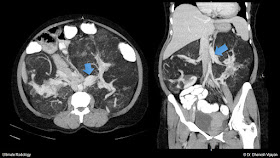
MIP (31mm) image on left shows the bilateral inferiorly oriented renal arteries, residual enhancing parenchyma of right kidney with fatty lesions. On the right, the axial post contrast images show the marked anterior displacement of the ascending colon (blue arrow) and descending colon (orange arrow).


3D VRT right lateral, frontal and left lateral views again shows the mass effect on colon which are seen markedly displaced anteriorly, against the anterior abdominal wall. Contrast filled small bowel loops are seen in RIF region.


3D VRT image showing the orientation of bilateral main renal arteries.


Delayed MIP images showing contrast excretion into the pelvicalyceal system of both kidneys. Right proximal ureter is marked by the orange arrow.


Delayed MIP axial and coronal images showing the right proximal ureter (orange arrow) and green arrows point to the opacification of pelvicalyceal system. No significant delay in contrast excretion was noted in this patient.


3D VRT of delayed excretory phase showing contrast within the pelvicalyceal system of both kidneys.


As expected renal veins showed moderate dilatation, with left renal vein measuring up to 17mm in diameter.


REFERENCES :

Indian J Nephrol. 2010 Apr; 20(2): 92–93. doi:  10.4103/0971-4065.65303, PMCID: PMC2931141, Renal replacement lipomatosis: A rare type of renal pseudotumor, N. A. Choh, M Jehangir, and S. A. Choh. https://www.ncbi.nlm.nih.gov/pmc/articles/PMC2931141/

Setty N, Uma K, Narvekar V N, Desai R S. Bilateral idiopathic replacement lipomatosis of the kidney with posterior mediastinal lipomatosis. Indian J Radiol Imaging [serial online] 2002 [cited 2017 Jun 21];12:251-2. Available from: http://www.ijri.org/text.asp?2002/12/2/251/28457