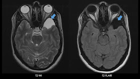
As its an extra-axial lesion it does buckles the grey-white interface and may also remodel / cause scalloping / thin adjacent bone.
It comprises of ~1% of all intracanial masses. Majority (~60%) are located in the middle cranial fossa, anterior the the temporal lobe, with posterior displacement of MCA. Less common sites of arachnoid cysts include CP angle, Suprasellar region, convexity and quadrigeminal cistern.
The major differential diagnosis will be an epidermoid cyst, porencephalic cyst, neurenteric cyst and neuroglial cysts. DWI and T2 FLAIR sequences are the most helpful in arriving at a diagnosis.
Epidermoid cysts show diffusion restriction. These lesions are only partially suppressed in T2 FLAIR images and looks 'dirty'. These are plastic lesions, which instead of displacing vessels engulf them and insinuate into the sulcal spaces.
A porencephalic cyst will usually have history of previous trauma or infarct. And these are usually surrounded by gliotic areas, not displaced cortex.
Neuroglial cysts are usually intra-axial.
Neurenteric cysts are usually seen in posterior fossa and they often contain proteinaceous fluid.
The following images depict the typical imaging findings of a middle cranial fossa arachnoid cyst.
In the anterior temporal location, it may be associated with temporal lobe hypoplasia. Above image shows complete homogenous supression of the signal of the arachnoid cyst in T2 FLAIR images.
Above image shows the lesion being isointense to CSF in T1 WI.
In DWI-ADC, contrast from an epidermoid cyst, the arachnoid cyst shows no diffusion restriction.
Galassi et al. had classified middle cranial fossa arachnoid cysts into 3 types long back in 1982, this classification is still being followed.
Type I cyst : Located in the Sylvian fissure, in the anterior temporal region, posterior to the sphenoid ridge, without any mass effect. These freely communicate with the subarachnoid space in Contrast CT cisternogram or in Phase Contrast MR evaluation.
Type II cyst : Located in the mid and proximal portions of Sylvian fissure, larger and rectangular in configuration. They communicate with subarachnoid space but slowly.
Type III cyst : Usually do not communicate with subarachnoid cyst, largest, lentiform in shape, will result in signficant mass effect and midline shift.
NOTE : A very large arachnoid cyst can show T2 slightly hypointense signal due to internal flow.


No comments:
Post a Comment